
Figure 6. Effects of IL36Ra or IL1Ra knockdowns on PMN and macrophage infiltration in B6 mouse cornea at 1 dpi.
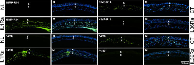
Mouse corneas were treated with control (CT, marked at right side), IL-1Ra (marked at left side), or IL-36Ra (marked at right side) siRNAs and inoculated with Pa as in Figure 3. Naïve corneas (NL, marked at left side) were used as negative control. The corneas were excised and processed for immunohistochemistry analysis at 1dpi. The 6 μ cryostat sections of the corneas were stained with antibody NIMP-R14 for neutrophils and F4/80 for macrophages. The images of infiltrated cells (Green) were merged with DAPI (blue nuclei) staining (M). E epithelium; S stroma. Two independent experiments were performed, 1 representative image for each condition is presented. Scale bar: 100 μm.