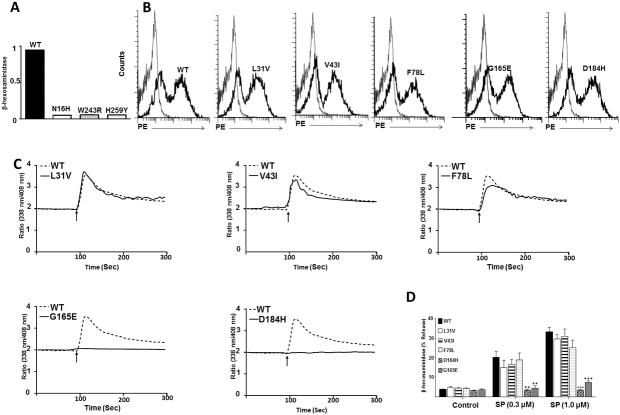
Figure 2. Effects of naturally occurring MRGPRX2 mutations (L31I, V43I, F78L, G165E and D184H) on cell surface expression, SP-induced Ca2+ mobilization and degranulation in stably transfected RBL cells.
(A): Cells were transfected with cDNA encoding wild-type (WT), N16H, W243R, or H253Y variant, transferred to 24 well plate and G418 was added to the culture medium 16 h after transfection. After 5 days, non-adherent cells were removed, adherent cells were lysed and total β-hexosaminidase content was determined. (B): Flow cytometry was performed with PE-anti-MRGPRX2 antibody to determine cell surface expression of WT and variants in stably transfected RBL cells. Representative histograms for WT/Variant (thick line) and control untransfected cells (thin line) are shown. (C): Cells expressing WT and MRGPRX2 variants were loaded with Indo-1 and intracellular Ca2+ mobilization in response to SP (1 μM) was determined. Data shown are representative of three independent experiments. (D): Cells were exposed to buffer (control) or SP (0. 3μM and 1 μM) for 30 min and β-hexosaminidase release was determined. All data points are expressed as mean ± SEM of three experiments performed in triplicate. Statistical significance was determined by two-tailed unpaired t-Test. *** indicates P value<.001, and ** indicates P value <.01.

