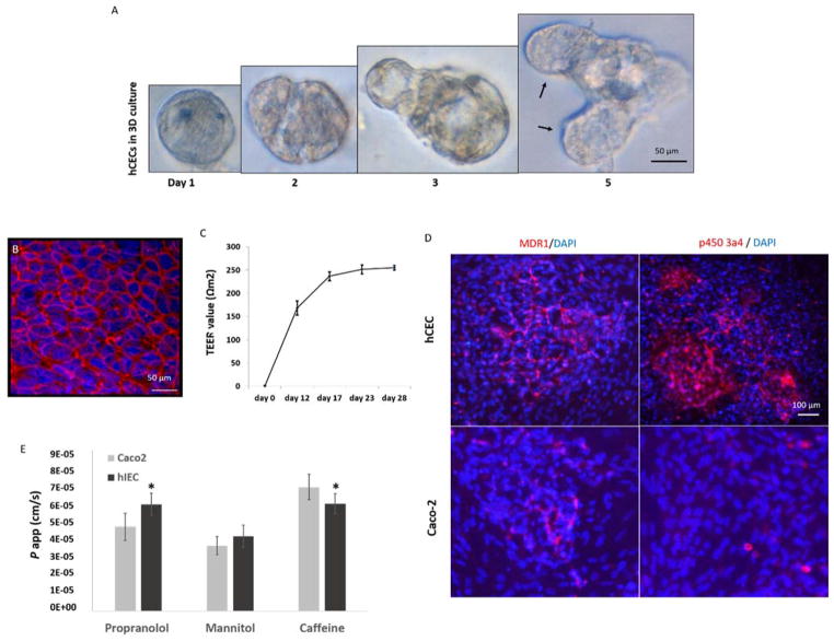
Figure 1. Culture of hIECs on 3D matrigel and transwell plates.
A. In matrigel cultures, individual hIECs progressively formed organoid-like structures (left to right). Black arrows denote micro-crypt domains formed with the multicellular cyst-like structure, which is the typical behavior of physiologically active intestinal stem cells grown in 3D culture. B. Tight junction formation on a polycarbonate membrane that was coated with Collagen-I; Red: ZO-1; Blue: DAPI (nuclear). Scale bars are present on the micrographs C. The hIECs derived tight junctions can be sustained for at least 28 days with a TEER value close to that in native human GI. D. Expression of MDR1 and P450 3A4 in hIECs and Caco2 cells, detected by immunostaining using antibodies against human MDR1 or P450 3A4; Red: MDR1 or P450 3A4; Blue: DAPI (nuclei). Scale bars are present on the micrographs E. hIECs or Caco2 cells were cultured on the polycarbonate membrane inserts of transwell plates for 28 days and the permeability coefficient (P app) of propranolol, mannitol or caffeine from the apical-to-basolateral (A to B) direction was measured as a correlation with intestinal drug absorption. Cell-free Transwell plates were used for negative control of non-specific binding. P app values are presented as mean (ng/ml) ± SEM (n = 3). Significant differences (P<0.05) between hIECs and Caco2 cells were observed in the absorption of propranolol and caffeine, noted with an asterisk. (by one-way ANOVA).

