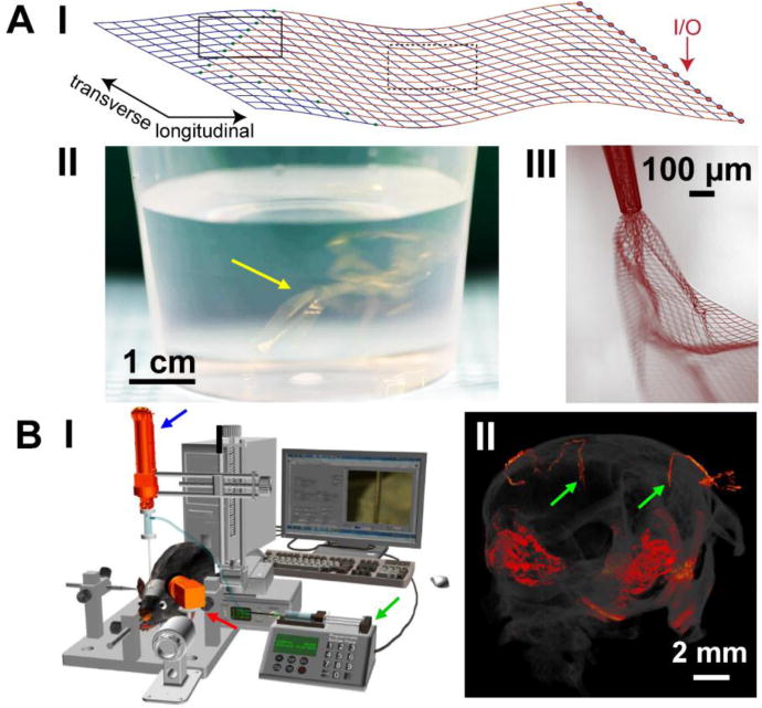
Figure 1.

Design and delivery of mesh electronics probes. (A) Unique structural and mechanical design of mesh electronics enables novel syringe-assisted delivery through a needle. I, Schematic of 16-channel mesh electronics, highlighting the recording electrodes (green dots in solid black box), metal interconnects (red lines in dashed black box) and I/O pads (red arrow). II, Photograph of a beaker with multiple 16-channel mesh electronics probes (yellow arrow) suspended in an aqueous saline solution; the golden color corresponds to light scattered and reflected from 10 μm wide, 100 nm thick metal interconnect lines. III, Bright-field microscope image taken in wide-field transmission mode showing partially ejected mesh electronics through a glass needle with an I.D. of 95 μm, exhibiting significant expansion and unfolding of mesh electronics in aqueous solution. (B) Controlled stereotaxic injection allows precisely targeted delivery of mesh electronics in the brain. I, Schematic of the semi-automated instrumentation for controlled injection of mesh electronics, highlighting a syringe pump for controlling the volumetric injection rate (green arrow), the motorized translation stage for controlling needle withdrawal (blue arrow), and the CCD camera for visualizing the mesh in the FoV (red arrow). II, Micro-CT image showing two fully extended mesh electronics probes (red linear structures highlighted by green arrows) within the brain following controlled injection. The other reddish areas correspond to the skull and mesh probe (I/O region) on the outer surface of the skull. Reproduced with permission from refs. 15 and 34.
