FIGURE 5.
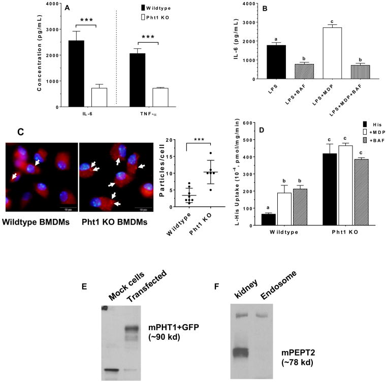
Effect of PHT1 on the LPS-MDP stimulated immune response, and uptake studies of MDP or L-histidine. (A) BMDMs from wildtype and Pht1 knockout mice were treated for 24 hr with 5 ng/mL LPS plus 10 μg/mL MDP, after which IL-6 and TNF-α concentrations in the cell culture media were measured. Data are expressed as mean ± SE (n=6) in which each experiment was performed in triplicate. Statistical differences between the two genotypes were determined by an unpaired (two-sample) t-test. ***p ≤ 0.001. (B) BMDMs from wildtype mice were treated for 24 hr with 5 ng/mL LPS alone, and in the presence of 1.0 μM bafilomycin A1 (BAF), 10 μg/mL MDP, or 10 μg/mL MDP plus 1 μM BAF. BMDMs were pretreated with BAF for 30 min prior to being co-incubated with MDP and/or LPS. IL-6 concentrations in the cell culture media were then measured. Data are expressed as mean ± SE (n=3) in which each experiment was performed in triplicate. Treatment groups with the same letter were not statistically different, as determined by ANOVA and Tukey’s test. (C) uptake of MDP-rhodamine in BMDMs prepared from wildtype and Pht1 knockout mice. MDP-rhodamine (red) was marked by arrows and the nuclei (blue) were stained by DAPI (100× magnification). Particle density is shown in the right-hand panel and expressed as mean ± SE (n=6–8 cells). Statistical differences between the two genotypes were determined by an unpaired (two-sample) t-test. ***p ≤ 0.001. (D) effect of potential inhibitors on the uptake of 1.0 μM [3H]histidine into endosome-enriched liver preparations from wildtype and Pht1 knockout mice. Endosomes were pretreated with MDP (1.0 mM) or BAF (1.0 μM) for 30 min prior to experimentation. Data are expressed as mean ± SE (n=3) in which each experiment was performed in triplicate. Treatment groups with the same letter were not statistically different, as determined by ANOVA and Tukey’s test. (E) fusion protein of GFP-mPHT1 (90 kd) was detected with an anti-GFP monoclonal antibody in the endosome of pcDNA3.1-mPht1/CT-GFP transfected HEK293 cells, but not in pcDNA3.1-CT-GFP transfected HEK293 (mock) cells. (F) expression of PEPT2 protein in mouse liver endosomes. One representative example is shown of three individual immunoblots.

