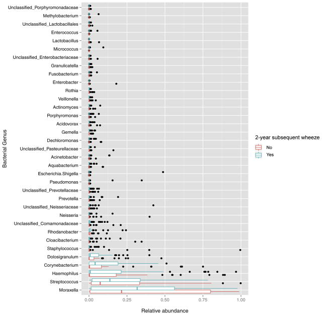
FIG. 2.
Boxplots of relative abundance of nasopharyngeal bacterial genera in infants with RSV ARI by 2-year subsequent wheeze. Within each sample, counts were normalized to simple proportions. The relative abundance of the 35 most abundant genera is shown; all other genera are not shown in this figure. The median (middle bar), third quartile (right-most bar), and first quartile (left-most bar) abundances are shown. Outliers are represented as dots.

