
FIGURE 1.
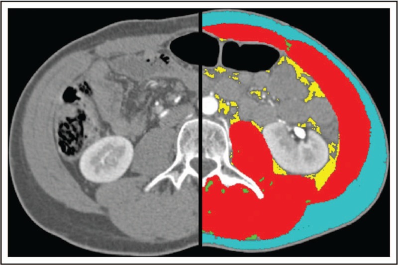
Cross-sectional image from abdominal computed tomography scan at the level of the third lumbar vertebra (L3), both unanalyzed (left) and analyzed (right) using SliceOmatic (TomoVision, Magog, Quebec, Canada). Red: muscle, green: intermuscular adipose tissue, yellow: visceral adipose tissue, and blue: subcutaneous adipose tissue.