
Figure 3.
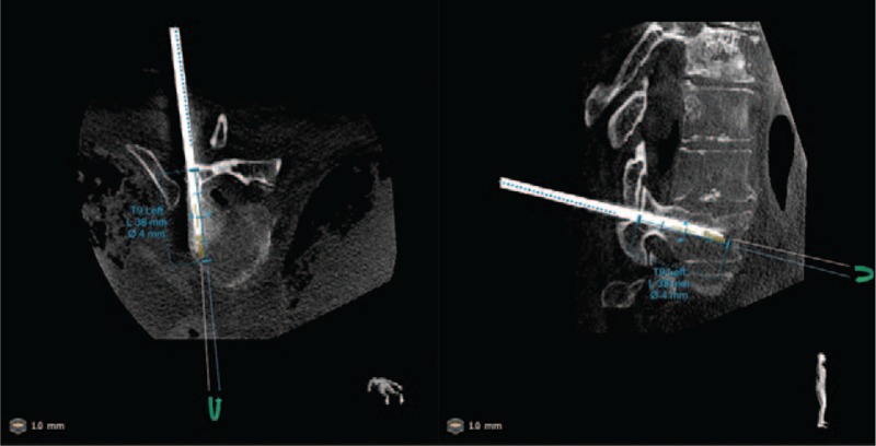
Postoperative cone beam computed tomography (CT) image showing axial and sagittal views of a Jamshidi needle at level T9 (left side) and the corresponding predefined path. In this case, a technical accuracy in the 3D space was 2 mm. Angulation error, being the angle between the predefined path (in blue) and the axis of symmetry of the needle, was measured on both planes and were 0.1 and 0.3 degrees on axial and sagittal planes, respectively.