
Figure 4.
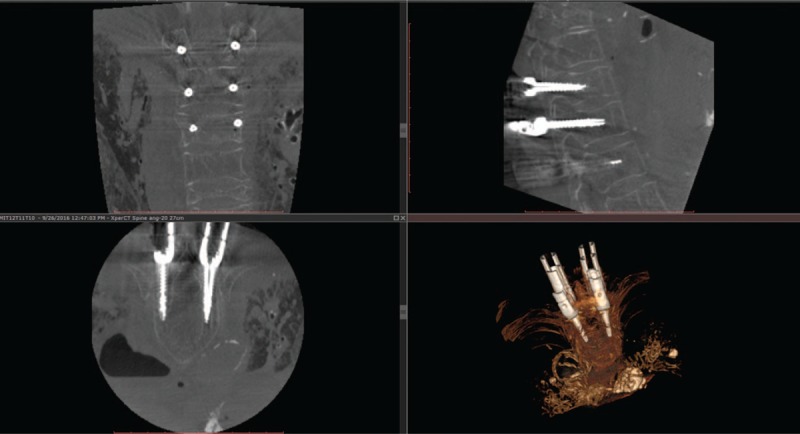
Example of a postoperative intraoperative images of six cannulated screws (bilateral on T10, T11, and T12) for minimally invasive spine (MIS) surgery on coronal (top left), sagittal (top right), and axial (bottom left) planes along with 3D volume rendering (bottom right). Note that there are hardly any metal artifacts despite the presence of six screws and the muscle dilators for each screw that was achieved thanks to the angulated acquisition scan. The axial slice depicts the screws inserted in T12 which both were graded as perfectly inside the pedicle (i.e., grade 0).