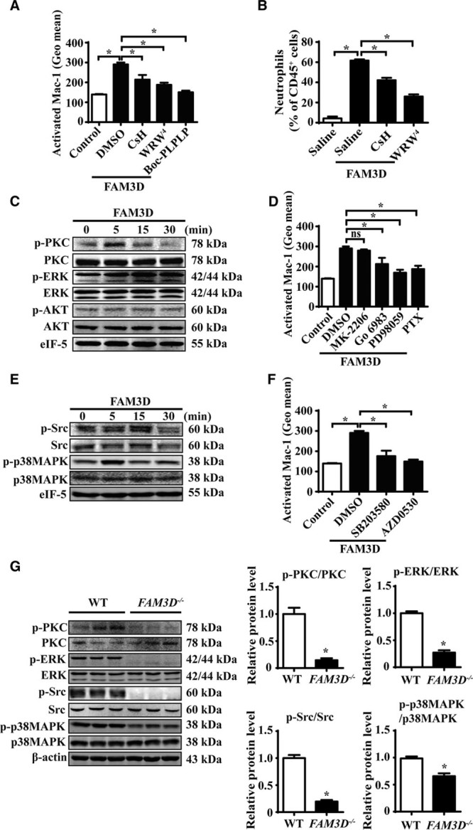
Figure 6.

FAM3D (family with sequence similarity 3, member D) induces Mac-1 (macrophage-1 antigen) activation via FPR (formyl peptide receptor)-Gi protein/β-arrestin signaling. A, Flow cytometric quantification of activated Mac-1 (CD11b) in human neutrophils pretreated with 10 ng/mL of Boc-PLPLP, 5 μmol/L of cyclosporine H (CsH), or 5 μmol/L of WRW4 for 2 hours followed by FAM3D (400 ng/mL) treatment for another 15 minutes. n=6. One-way analysis of variance (ANOVA) followed by Tukey’s test for multiple comparisons, *P<0.05. B, C57BL/6 mice (11–12 weeks old) underwent the intraperitoneal injection of CsH (10 μg/30 g body weight) or WRW4 (10 μg/30 g body weight) at 1 hour prior to FAM3D induction. Then FAM3D (10 μg/30 g body weight) was injected into peritoneal cavity for neutrophil recruitment in 6 hours. The percentage of neutrophils in CD45+ peritoneal cells was analyzed as CD45+CD11b+Ly6G+ by flow cytometry. n=7. One-way ANOVA followed by Tukey’s test for multiple comparisons, *P<0.05. C, Representative Western blot analysis of PKC (protein kinase C), ERK (extracellular regulated MAP kinase), and AKT (PKB, protein kinase B) activation in human neutrophils treated with 400 ng/mL FAM3D for various periods. D, Human neutrophils were pretreated with 20 μmol/L of MK-2206, 100 nmol/L of Go6983, 10 μmol/L of PD98059 or 10 ng/mL of PTX for 2 hours, followed by FAM3D (400 ng/mL) treatment for another 15 minutes, and the activation of Mac-1 (CD11b) was analyzed by flow cytometric assay. n=6. One-way analysis of variance (ANOVA) followed by Tukey’s test for multiple comparisons, *P<0.05, ns, no significance. E, Representative Western blot analysis of Src and p38MAPK (mitogen-activated kinase-like protein) activation in human neutrophils treated with 400 ng/mL FAM3D for various periods. F, Human neutrophils were pretreated with 10 μmol/L of SB203580 or 5 μmol/L of AZD0530, respectively, for 2 hours followed by FAM3D (400 ng/mL) treatment for another 15 minutes; the activation of Mac-1 was analyzed by flow cytometric assay. n=6. One-way ANOVA followed by Tukey’s test for multiple comparisons, *P<0.05. G, Representative Western blot analysis and quantification of PKC, ERK, Src, and p38MAPK in peripheral blood neutrophils isolated from wild-type (WT; n=6) and FAM3D−/− (n=6) mice treated with elastase for 7 days. Unpaired Student t test, *P<0.05. HUVECs indicates human umbilical vein endothelial cells; PTX, pertussis toxin; and TNF-α, tumor necrosis factor α.
