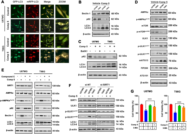
Fig. 3. Comp 5 induces autophagy via AMPK-mTOR-ULK complex axis in GBM cells.
a U87MG and T98G cells were transfected with GFP-mRFP-LC3 plasmid, after co-incubation with 10 μM Comp 5, the GFP-LC3 puncta were observed by fluorescence microscope. Scale bar = 20 μm. b U87MG and T98G cells were treated with 10 μM Comp 5 for 24 h, then the expression levels of Beclin-1, p62, and LC3 were determined by western blot. c U87MG and T98G cells were treated with Comp 5 in the presence or absence of BafA1, then the accumulation of p62 and LC3 were detected by western blot. d U87MG and T98G cells were treated with 10 μM Comp 5 for 24 h, then the expression levels of AMPKα, p-AMPKαthr172, mTOR, p-mTORser2448, ULK1, p-ULK1ser317, p-ULK1ser555, mATG13, FIP200, and ATG101 were determined by western blot. e U87MG and T98G cells were treated with Comp 5 in the presence or absence of Compound C, then the expression levels of SIRT1, AMPKα, p-AMPKαthr172, p62, Beclin-1, and LC3 were detected by western blot. f U87MG and T98G cells were transfected with SIRT1 siRNA for indicated time and treated with Comp 5 for additional 24 h. The expression levels of SIRT1, ULK1, p-ULK1ser317, p-ULK1ser555, and LC3 were determined by western blot. β-actin was used as a loading control. g U87MG and T98G cells were treated with 10 μM Comp 5 in the presence or absence of 3-MA (1 mM) for 24 h. Then, cell viabilities were detected by MTT assay. ***P < 0.001 vs. Comp 5-treated group

