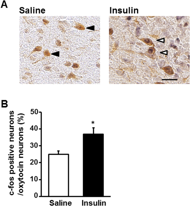
Figure 2.

Effect of insulin on c-Fos expression in oxytocin-IR neurons. (A) Representative pictures depicting dual immunostaining for c-Fos and oxytocin in PVN after injection of saline or insulin (100 μU/2 μl). Black arrowheads indicate oxytocin-IR neurons. White arrowheads indicate the neurons IR to both oxytocin and c-Fos. Scale bar: 30 μm. (B) Incidence of c-Fos-IR neurons in oxytocin-IR neurons. n = 5. *p < 0.05 vs. saline by one-way ANOVA followed by Tukey’s test. Error bars are SEM.
