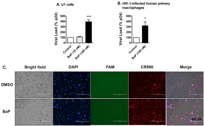
Figure 1.
Chronic treatment of BaP induces HIV-1 replication and apoptotic DNA damage in HIV-1-infected macrophages. (A) The U1 cells were treated with 10 nM and 100 nM BaP for seven days. After the BaP treatment, the U1 cells were stimulated with 100 nM of Phorbol 12-myristate 13-acetate (PMA) to produce HIV-1. Supernatants were collected after two days of differentiation, which were used for the p24 ELISA assay to assess the viral load. The chronic (7 days) treatment of BaP (100 nM) significantly increased the viral replication in U1 cells, while the 10-fold lower concentration did not have any significant effect. The data is displayed as mean ± SEM (n = 6), calculated as a percentage of the control. (B) HIV-infected human primary macrophages were treated with BaP (100 nM) for 3 days. The supernatant was collected thereafter and used for the p24 ELISA assay to assess the viral load. The acute (3 days) treatment of BaP (100 nM) significantly increased the viral replication in HIV-1-infected primary macrophages. The data is displayed as mean ± SEM (n = 4). For calculating the viral load, we subtracted the nonspecific background reading from the actual absorbance values. Since the residual viral load varies from experiment to experiment in U1 cells, we normalized the control values for each experiment to 100% and calculated the values for the treated, as the percentage of the control. The statistical significance was calculated at *p ≤ 0.05, where *** represents p ≤ 0.0005, compared with the control group. (C) The apoptotic DNA damage assay was performed on the treated cells. DAPI, FAM and CR590 stained nucleus (blue), apoptotic DNA damage with DNase Type II ends (green) and Type I ends (red) respectively. A higher signal for CR590 is visible in the fluorescent images, indicating apoptotic DNA fragmentation with DNase Type I ends in the infected human primary macrophages after BaP (100 nM) exposure for 3 days. DNA fragmentation with DNase Type II ends (green) was not visible in either the control or the treated cells. Therefore, the images indicate that BaP (100 nM) induces DNA fragmentation during the early phase of apoptosis in the HIV-1-infected human primary macrophages.

