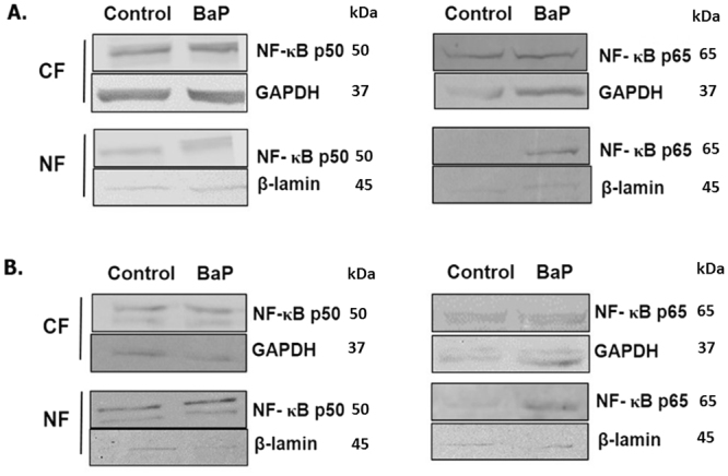
Figure 6.

Translocation of NF-κB subunits from cytoplasm to nucleus upon BaP exposure. U1 cells were treated with BaP 100 nm (A) and 1 µM (B) for 7 days and 3 days, respectively. After the treatment, proteins from the cytoplasm and nucleus were extracted from the cells. Western blot was run to determine the expression of the NF-κB p50 and p65 subunits in the proteins in cytosolic fraction (CF) and nuclear fraction (NF). GAPDH and β-lamin were used as loading controls for the cytoplasmic and nuclear proteins, respectively. The blots are representative of at least three independent experiments. There is not much difference in the expression of NF-κB p50 and p65 between the control and the BaP-treated cells in the cytoplasmic fraction. However, there is a clear increase in the expression of both the subunits in the nuclear fraction of acutely or chronically BaP-treated cells compared to the control group.
