Figure 2.

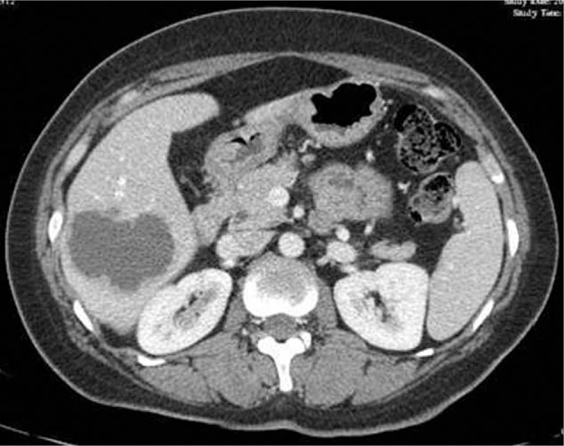
Computed tomography presenting a low density and heterogeneous lesion taking irregular wall and incompletely septa with strong contrast enhancement.

Computed tomography presenting a low density and heterogeneous lesion taking irregular wall and incompletely septa with strong contrast enhancement.