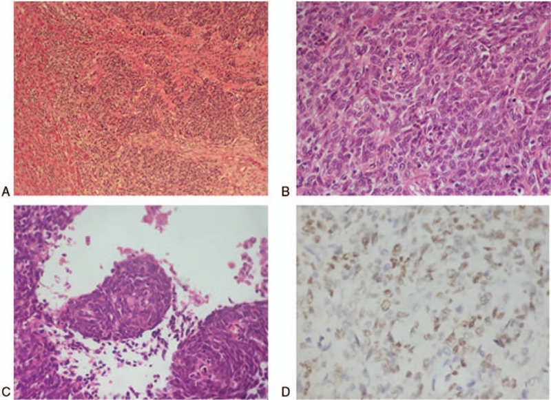
Figure 3.

Resected segment biopsies. (A) Tumor with ill-defined boundary had the feature of infiltrative growth (hematoxylin and eosin stain, ×40). (B) Poorly differentiated cells presenting round or ovoid in shape consistent with primary nasopharyngeal carcinoma (×400). (C) Squamous eddy can be seen indicating squamous cells differentiation (×400). (D) Tumor cells were Epstein–Barr ribonucleic acid positive in situ hybridization (×400).
