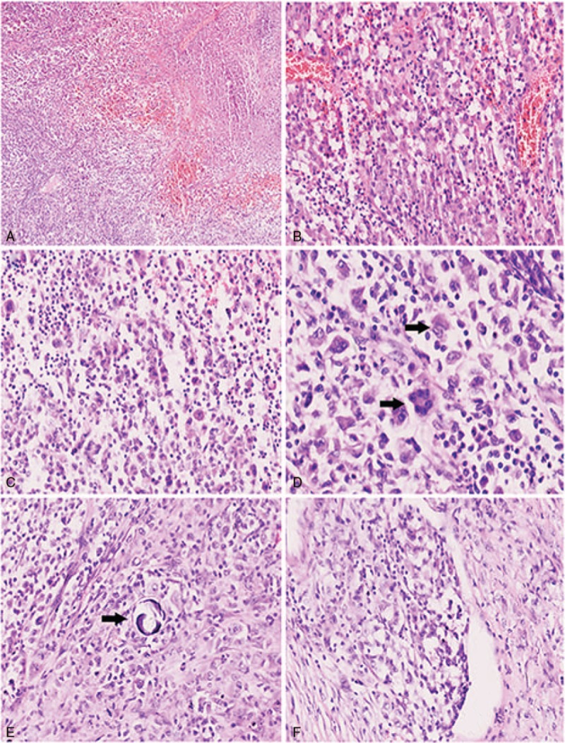
Figure 1.

Numerous tumor cells with easily identified foci of necrosis (A, 40×); the background consisted of inflammatory cells, hemorrhage and congested vessels, tumor cells were highly pleomorphic and exhibited eosinophilic cytoplasm (B, 200×); neoplastic cells show loose arrangement (C, 200×); the nuclei were medium to large in size with a distinct oval to kidney shape (black arrow), thickened nuclear membranes and granular chromatin, polynuclear tumor cells could be identified (black arrow, D, 400×); calcification rarely present (E, 200×); vascular invasion was easily identified (F, 200×).
