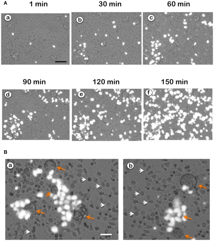
Figure 2.
Neutrophils in touch with Entamoeba histolytica release NETs. (A) Human neutrophils were stimulated with E. histolytica in the presence of SYTOX® Green. Live cell images were captured at different times with a fluorescence inverted microscope. External DNA fluorescence appears bright white in the pictures. Scale bar is 50 μm. (B) NETs formation was induced only in neutrophils that were in direct contact with E. histolytica trophozoites (orange arrows). The NETs were produced around the amoebas and progressively covered the parasites. Neutrophils that were not in contact with amoebas (white arrow heads) did not release DNA fibers and never became SYTOX® Green-positive. Scale bar is 25 μm.

