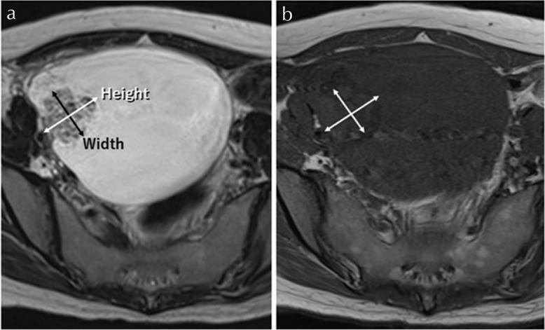
Fig. 3.

Mural nodules with malignant features on MR imaging. Clear cell carcinoma, stage I C3, in 39-year-old woman. (a) On axial T2-weighted images (T2WI), the mass demonstrates high-signal intensity with a mural nodule on the anterior-right sided wall of the cyst. (b) The signal intensity of the mass on axial T1-weighted images (T1WI) indicated homogenous low signal. “Height” was 2.9 cm and “Width” was 2.5 cm. “Height-Width ratio (HWR)” indicated 1.16.
