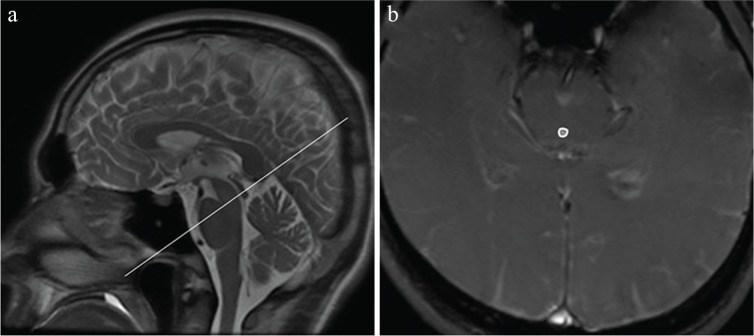
Fig. 1.
Sample anatomic and velocity-encoded images. (a) Midline sagittal T2 weighted magnetic resonance scout image; the line indicates the location of the plane through the midcollicular level at the aqueduct, defining the plane of measurement for the cerebrospinal fluid flow velocity. (b) Portions of the velocity-encoded image with a region of interest placed in the aqueduct of the midbrain.

