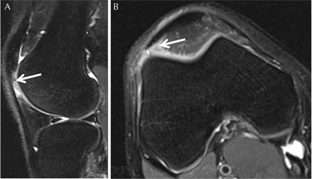
Fig. 5.
Superolateral infrapatellar fat pad (Hoffa fat pad) impingement in a 37-year-old man. (A) Sagittal and (B) axial fat-suppressed proton-density weighted MR images demonstrate high signal in the superolateral aspect of Hoffa fat pad between the lateral aspect of the patellar tendon and the lateral femoral condyle (arrow). Note short distance between the lateral aspect of the patellar tendon and the lateral femoral condyle (solid line). Modified Insall-Salvati ratio was within normal limits in this case, although the tibial tubercle - trochlear groove distance (not shown) at 20 mm was consistent with lateral patellar shift.

