Fig. 6.
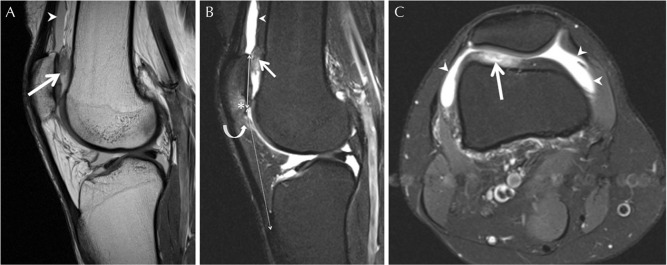
Prefemoral fat pad impingement in a 42-year-old woman with anterior pain of the right knee. (A) Sagittal proton-density MR image. (B) Sagittal fat-suppressed proton-density MR image. (C) Axial fat-suppressed proton-density MR image. Enlargement and signal change of the inferolateral aspect of the prefemoral fat pad (low signal on proton-density and high signal on fat-suppressed proton-density, as shown by solid arrows) with bulging and mass effect toward the suprapatellar pouch, and associated joint knee effusion (arrowheads). Note associated full-thickness cartilage defect and bone marrow lesion of the lower patella (asterisk), and edema of the superolateral aspect of the infrapatellar fat pad (Hoffa fat pad) (curved arrow) consistent with associated patellofemoral maltracking. Note high riding patella with modified Insall Salvati ratio: distance between distal patellar articular surface and distal patellar tendon insertion (dashed double end arrow)/patellar length (solid double end arrow) measured at 2.4 (greater than 2).

