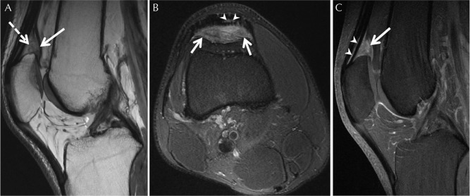
Fig. 9.
Suprapatellar fat impingement in a 29-year-old woman with chronic anterior knee pain. (A) Sagittal proton-density MR image shows enlargement and mass effect of the suprapatellar fat pad (solid arrow). Note low signal intensity within the fat pad in comparison with other adipose structures in the knee joint that are indicative of inflammation (dashed arrows). (B) Axial fat-suppressed proton-density image reveals high signal intensity and enlargement of the fat pad extending into the endotenon fat of the distal quadriceps entheses (arrowheads). (C) Sagittal fat-suppressed gadolinium-chelate contrast medium-enhanced T1-weighted image shows diffuse homogenous contrast enhancement within the suprapatellar fat pad extending into the endotenon fat within the distal quadriceps enthesis (arrowheads).

