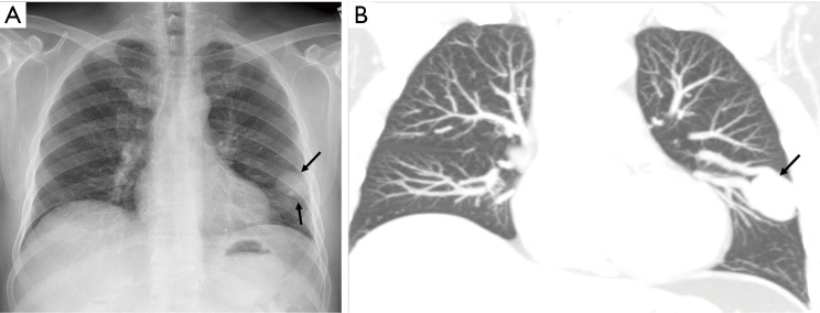
Figure 10.
Frontal radiograph of the chest (A) in a 61-year-old man with HHT showing round soft tissue non-calcified nodule (arrows) in the left mid zone. Corresponding coronal CT image (B) shows 3.5 cm aneurysmal connection along with feeding artery and draining vein in lingula lobe (arrow) related to pulmonary arteriovenous malformation (PAVM).

