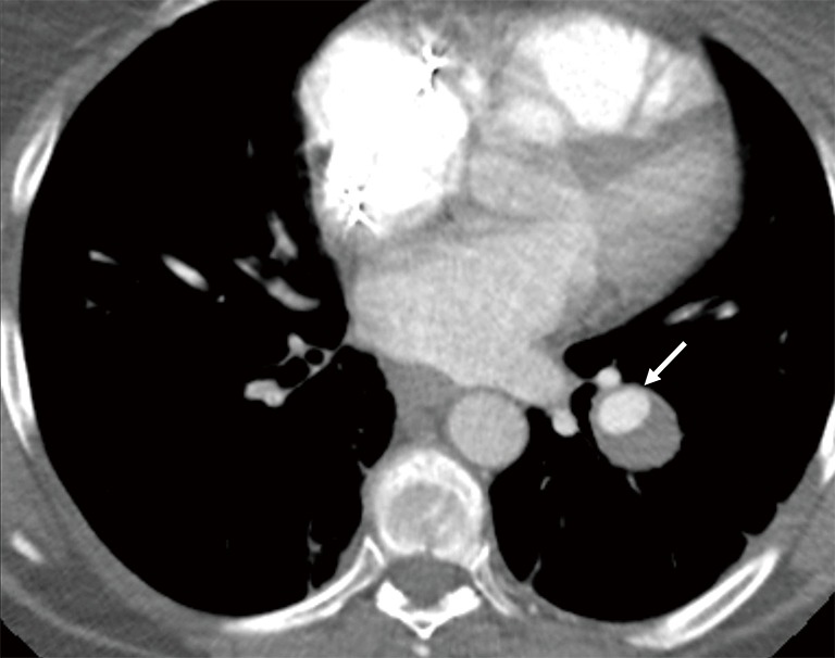
Figure 13.

Axial contrast enhanced CT image in a 50-year-old man with left lower lobe pulmonary artery aneurysm with partial thrombosis (arrow) connecting with branch pulmonary artery. Note lack of connecting draining vein which is typical feature of PAVM.
