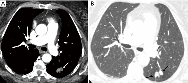
Figure 15.
Bronchocele. Axial contrast enhanced CT image in soft tissue (A) and lung window (B) in a 65-year-old man shows non-enhancing nodular branching soft tissue low attenuation lesion in left lower lobe superior segment related to bronchocele (arrows) due to localized bronchial atresia and hyperlucency in adjacent lung. Note lack of feeding artery and draining vein which are typical feature of PAVM.

