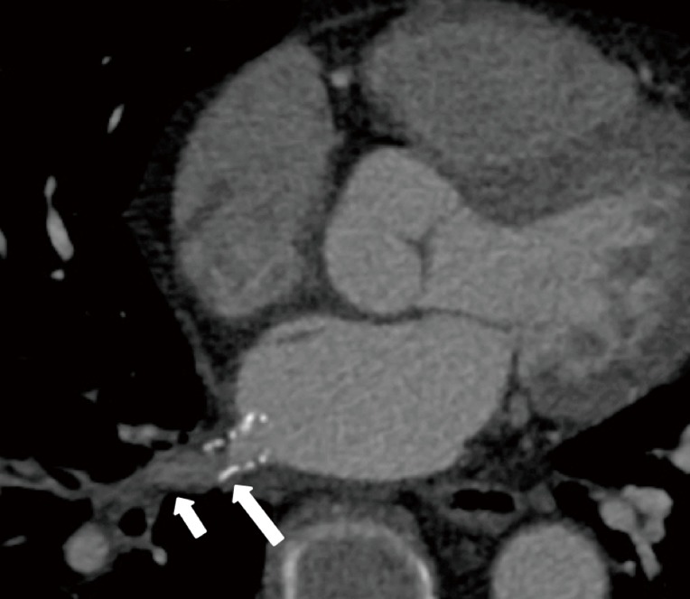
Figure 10.

In-stent restenosis after percutaneous stenting of a right inferior pulmonary vein stenosis secondary to fibrosing mediastinitis. Axial reconstructed CT image demonstrates hypoattenuating material in the distal half of the stent (long arrow), suggestive of intimal proliferation, and poor contrast opacification of the pulmonary vein segment distal to the stent (short arrow), suggesting in-stent restenosis.
