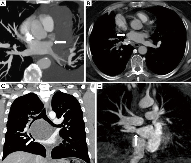
Figure 3.
Acquired pulmonary vein stenosis in adults secondary to lymphoma, fibrosing mediastinitis, bronchogenic cyst, and percutaneous radiofrequency catheter ablation for atrial fibrillation. (A) Axial reconstructed maximal intensity projection CT image shows lymphoma demonstrating mass effect on the left superior pulmonary vein with resultant stenosis (arrow); (B) axial reconstructed CT image shows fibrosing mediastinitis demonstrating mass effect on the right superior pulmonary vein with resultant stenosis (arrow); (C) coronal CT image shows bronchogenic cyst demonstrating mass effect on the right superior pulmonary vein with resultant stenosis (arrow); (D) coronal reconstructed maximal intensity projection MR image shows right inferior pulmonary vein stenosis after percutaneous radiofrequency catheter ablation for atrial fibrillation (arrow).

