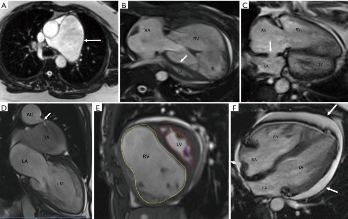
Figure 7.
Magnetic resonance imaging. (A) Axial steady state free precession image (SSFP) shows severe dilation of the main pulmonary artery (arrow). (B) Axial SSFP image shows severe dilation and hypertrophy of the right ventricle (RV), with decreased size of the left ventricle (LV). The interventricular septum is flattened and bowed to the left (arrow). (C) Axial SSFP image shows a moderate-sized secundum type of atrial septal defect (arrow). Note the enlargement of the right ventricle and right atrium with flattening of the ventricular septum. (D) Two-chamber SSFP image shows a patent ductus arteriosus (arrow) connecting the aortic arch (AO) and a dilated left pulmonary artery (PA). (E) Short axis SSFP MRI image shows quantification of the RV by drawing endocardial contour in the end-diastolic image (yellow contour) and quantification of the LV by an endocardial contour in the end-diastolic image (red contour). (F) Axial SSFP image in a patient with pulmonary hypertension shows a moderate-sized circumferential pericardial effusion (arrows). Note also the dilated and hypertrophied right ventricle (RV). RA, right atrium; LA, left atrium.

