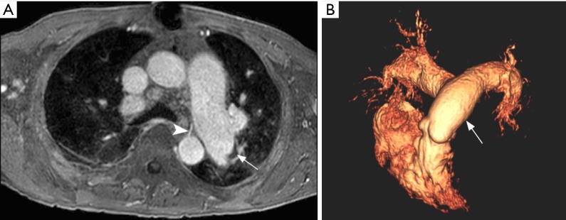
Figure 11.
Magnetic resonance pulmonary angiography (MRPA). Axial post contrast fat-suppressed spoiled gradient recalled echo (A) shows left pulmonary artery dilation with a subtle eccentric crescent-shaped thrombus (arrow). Note a prominent bronchial artery arising from the descending aorta (arrowhead). Tridimensional volume rendered reconstruction (B) showing dilation of the main pulmonary artery (arrowhead) and central branches.

