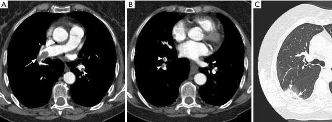
Figure 13.
CT pulmonary angiography (CTPA) images in acute pulmonary embolism. Axial CTPA images (A and B) show partial filling defects, centrally located in the right pulmonary artery (arrow) and eccentric in segmental branches (arrowheads). Note acute angle between thrombi and vessel walls. Axial lung window (C) shows “reversed halo sign” in the right lower lobe (arrows), a nonspecific sign of pulmonary infarction.

