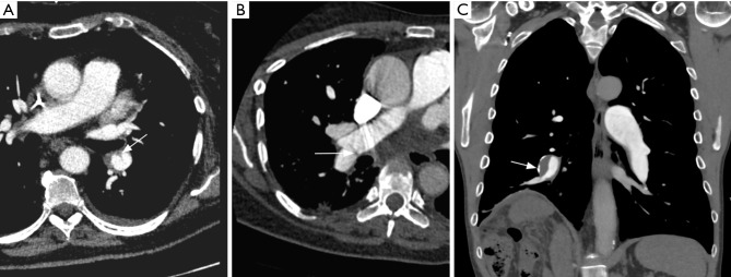
Figure 5.
CT pulmonary angiography appearances of chronic thrombi in different patients. Axial images (A) and (B) show webs (arrows), reflecting non-resolved thrombi and partial recanalization. Coronal image (C) shows an eccentric thrombus in right interlobar pulmonary artery with calcified mural thickening (arrow).

