Fig. 5.
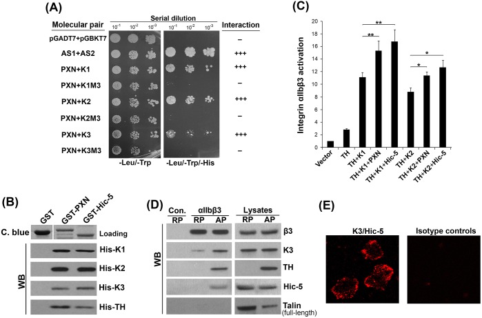
PXN family members interact with the kindlin family members and support integrin αIIbβ3 activation. (A) The interaction of PXN with different kindlins and their M3 mutants were evaluated using the Matchmaker™ Gold yeast two-hybrid system. (B) The interactions of PXN/Hic-5 with kindlins and TH were evaluated by pull-down assays. The precipitates were analyzed by western blotting (WB). Meanwhile, the loaded GST proteins on the beads was evaluated by Coomassie Blue (C. blue) staining. (C) As indicated, Flag–PXN (PXN) and Flag–Hic-5 (Hic-5) were each co-expressed with DsRed-fused TH (TH) and EGFP-fused kindlins (K1 and K2) in CHO-αIIbβ3 cells. Integrin αIIbβ3 activation in the positively transfected cells was measured by the PAC-1 binding assay. The results represent mean±s.d. *P<0.05, **P<0.01. (D) Washed human platelets were activated with TRAP-6 (7 μM, 15 min) or kept under resting conditions. Activated platelets (AP) and resting platelets (RP) were lysed and incubated with either control IgG or an antibody against αIIbβ3, in the presence of protein A/G agarose beads. After incubation for 6 h at 4°C, the beads were washed and the precipitated proteins on the beads analyzed by western blotting (WB). (E) Proximity ligation assay (PLA). Washed human platelets were spread on fibrinogen, PLA was performed and the proximity of Hic-5 and kindlin-3 was detected by confocal microscopy.

