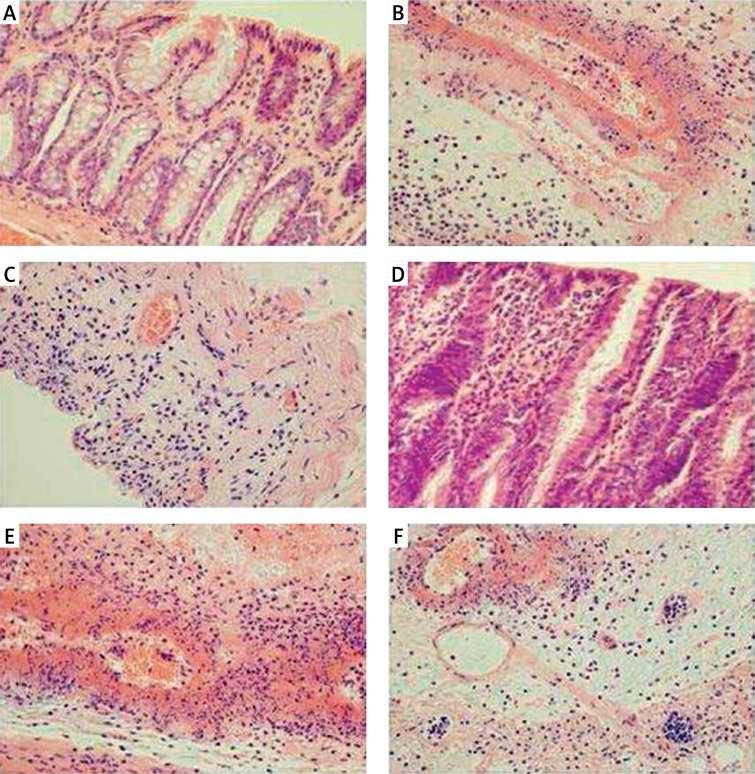
Figures 2.
A – Representative microscopic image of colonic mucosa observed 1 h after enema with saline in saline-pretreated control rats. B – Representative microscopic image of colonic mucosa observed 1 h after enema with acetic acid solution in saline-pretreated rats. C – Representative microscopic image of colonic mucosa observed 1 h after enema with acetic acid solution in rats pretreated with obestatin given at a dose of 8 nmol/kg/ dose. D – Representative microscopic image of colonic mucosa observed 24 h after enema with saline in saline-pretreated control rats. E – Representative microscopic image of colonic mucosa observed 24 h after enema with acetic acid solution in saline-pretreated rats. F – Representative microscopic image of colonic mucosa observed 24 h after enema with acetic acid solution in rats pretreated with obestatin given at a dose of 8 nmol/kg/dose. Hematoxylin-eosin stain. Original magnification 400×

