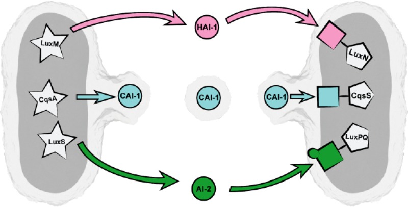
FIG 7.
Model of trafficking of CAI-1 via OMVs between V. harveyi cells. Due to its high lipophilicity, CAI-1 remains trapped in the outer membrane of V. harveyi cells and gets incorporated into OMVs. In this form, CAI-1 can be easily transported between cells and can activate the QS cascade in neighboring cells upon recognition by its specific receptor CqsS. The other QS signaling molecules of V. harveyi, AI-1 and HAI-1, are supposed to be freely diffusible across the cell envelope and are sensed by their specific receptors LuxPQ and LuxN, respectively, which are located in the inner membrane. The AIs HAI-1, CAI-1, and AI-2 are depicted with circles. AI synthases are depicted in stars in the cytoplasm. The AI-specific receptors LuxN, CqsS, and LuxPQ are depicted by rectangles in pink, blue, or green, respectively, connected to pentagons. The image was created by Christoph Hohmann (Nanosystems Initiative Munich).

