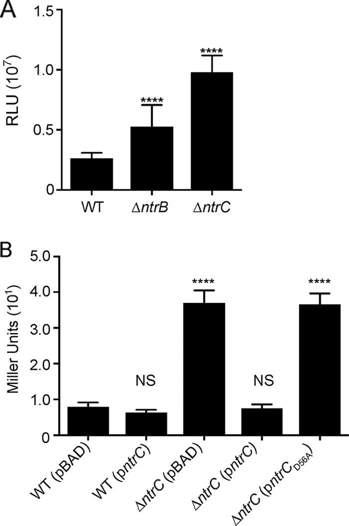
FIG 6.

NtrB and aspartate 56 of NtrC contribute to vpsL repression. (A) Expression of PvpsL-luxCADBE (pFY_3406) in the WT, ΔntrB, and ΔntrC strains. The graph represents the averages and standard deviations of RLU obtained from at least three technical replicates from four independent biological samples. RLU are reported in luminescence counts per minute per milliliter per OD600. Expression levels of vpsL in the ΔntrB and ΔntrC strains were compared to that in the WT. (B) β-Galactosidase assay of PvpsL-lacZ reporter strains containing either the empty vector (pBAD) or a vector expressing ntrC (pBAD-ntrC) or ntrCD56A (pBAD-ntrCD56A) under the control of an arabinose-inducible promoter. Cells were grown in LB medium supplemented with 0.1% arabinose to mid-exponential phase. The graph represents the averages and standard deviations of Miller units obtained from four technical replicates from two biological replicates. The expression levels of vpsL in all strains were compared to that in the WT (pBAD). ANOVA followed by Dunnett's test was performed for multiple-comparison analysis. ****, adjusted P value of <0.0001; NS, not significant.
