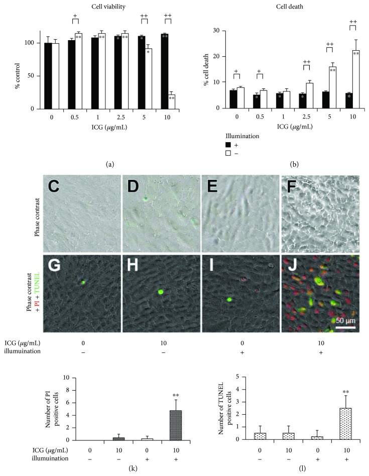
Figure 2.
Phototoxicity of ICG exposed to RPE cells under illumination. Cells were cultured with 0 to 10 μg/mL ICG in the dark or under illumination for 24 hours. (a) Cell viability and (b) cell death rate are shown. Cell viability of each culture measured by MTS assay was normalized to that of 0 μg/mL ICG. (c–f) Phase-contrast micrographs. (g, h) Fluorescence micrographs of PI staining and TUNEL. Quantitation of PI-positive cells (k) and TUNEL-positive cells (l) in 10,000 μm2. At least four squares of each experiment were analyzed. Dunnett's test: ∗P < 0.05 and ∗∗P < 0.01, unpaired Student's t-test: +P < 0.05 and ++P < 0.01.

