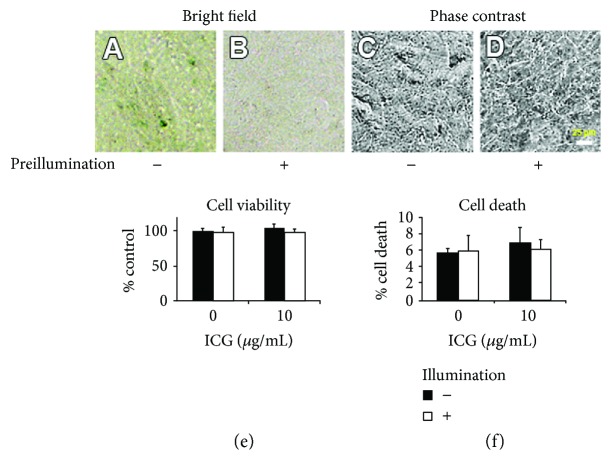
Figure 5.
Staining ability and cytotoxicity of the preincubated medium with ICG in the dark or under illumination. (a, b) Bright-field micrographs and (c, d) phase-contrast micrographs of cultures in the preincubated medium containing 10 μg/mL ICG in the dark or under illumination after 24 hours. (e, f) Cell viability and cell death rate in the preincubated medium containing 0 or 10 μg/mL ICG after a 24-hour culture. Cell viability of each culture was normalized to that of culture in the preincubated medium with 0 μg/mL ICG in the dark. There was no significant difference in cell viability and cell death rate among all conditions of cultures.

