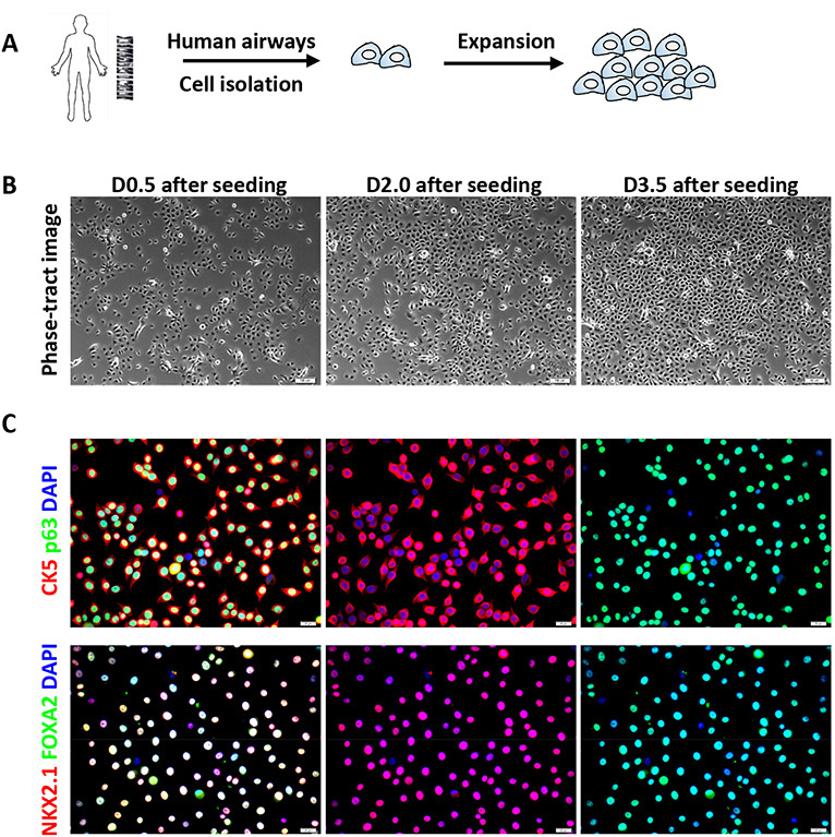
Figure 1. Serial expansion and immunofluorescence characterization of human airway basal cells.

A. Schematic of serial expansion of human airway basal cells. B. Phase contrast images of the human basal cells at various days after cell seeding (initial seeding density is 10-15%). Scale bars = 50 μm. C. Immunofluorescence of basal stem cell markers (CK5 and p63) and airway-specific transcription factors (NKX2.1 and FOXA2) on cultured airway basal cells (Passage 6). Scale bars = 20 μm.
