
Figure 1.
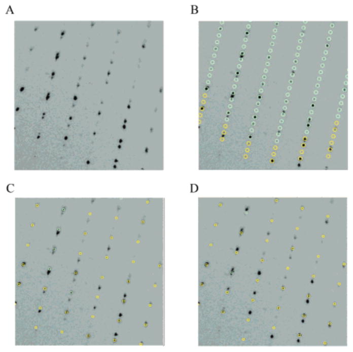
TbpA twinned diffraction pattern. (A) Section of the diffraction pattern showing two lattices. (B) Autoindexing of the diffraction pattern showing a monoclinic cell with unit cell parameters a = 69.4 Å, b = 119.8 Å, c = 274.2 Å, and β = 93.9°. Closer inspection of (A) and (B) reveals that some predictions have no intensities and that some spots are poorly predicted. Once the nonmerohedral twinning is taken into account, the same diffraction pattern is indexed with c = 91.9 Å. (C) Same diffraction pattern indexed with the correct unit cell depicting one twin domain. (D) Diffraction pattern indexed with the second twin domain.