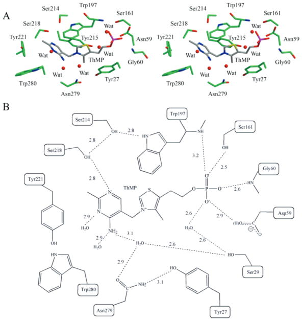
Figure 4.
TbpA ligand binding site. (A) Stereoview of the binding site with the amino acid side chains and ThMP shown in ball-and-stick. The binding site is colored by atom with green for carbon on the protein and gray for carbon in ThMP, blue for nitrogen, yellow for sulfur, red for oxygen, and magenta for phosphorus. (B) Schematic representation of the binding site showing ThMP key stacking interactions and hydrogen bonding to TbpA.

