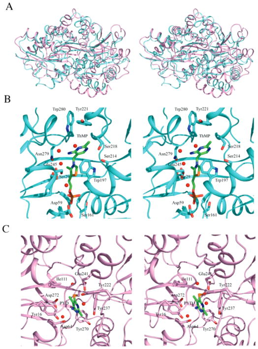
Figure 7.
Structural comparisons to thiaminase-I. (A) Stereoview of superposition of TbpA, shown in cyan, to thiaminase-I, shown in pink. (B) Stereoview of TbpA binding site. ThMP is shown as ball-and-stick colored by atom with green for carbon, blue for nitrogen, yellow for sulfur, red for oxygen, and magenta for phosphorus. Binding site side chains are shown in ball-and-stick with cyan for carbon, red for oxygen, and blue for nitrogen. Water molecules are shown as red spheres. (C) Stereoview of the thiaminase-1 binding site. The inhibitor PYD is covalently bound to Cys113 and is shown as ball-and-stick colored by atom with green for carbon, blue for nitrogen, and orange for sulfur. The binding site side chains are shown in ball-and-stick with pink for carbon and red for oxygen.

