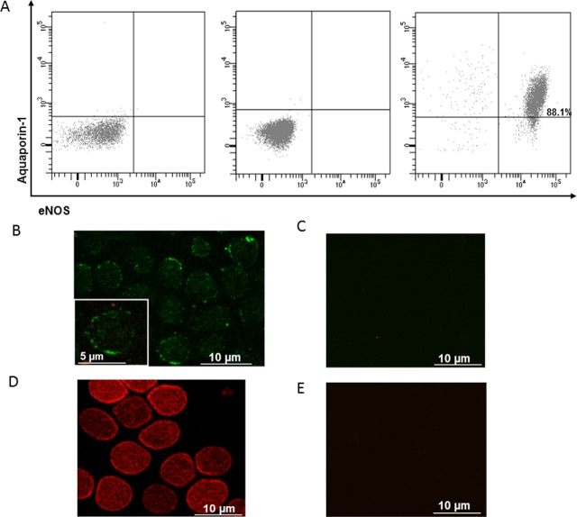
Fig 4. Human RBCs express eNOS proteins.
A. Representative flow cytometric two parameter dot plot of isolated RBCs from a healthy subject co-stained with primary anti-Aquaporin-1 and secondary Alexa Fluor-488-conjugated anti-IgG antibodies; and primary anti-eNOS and secondary Alexa Fluor-647-conjugated anti-IgG antibodies. Panel on the left shows co-staining only with conjugated secondary antibodies, panel in the middle shows staining only with mouse isotype IgG1,k Alexa-647-conjugated (negative controls). Panel on the right shows the identification of RBCs as eNOS–positive and Aquaporin1-positive events in the upper right quadrant. The percentage of RBCs double positive is indicated in the upper right quadrant. (B-E) Representative images of eNOS (B) and AQP1 (D) detection in healthy human RBCs using immunofluorescence microscopy. As negative controls, RBCs were stained only with conjugated secondary antibodies (Alexa Fluor-488 anti-mouse IgG for eNOS (C) and Alexa Fluor-568 anti-rabbit IgG for AQP1 (D)).

