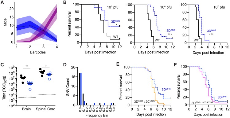
Fig 4. In vivo phenotype of WT and 3DG64S.
(A) Maximum likelihood optimization of a simple binomial model (see S1 Text Model 2) estimated an average inoculum to CNS bottleneck size of 2.67 (lambda 2.44; 95% CI 1.39–3.82) based on experimental data for 4 barcoded poliovirus populations [49]. Shown are outputs of 10,000 simulations of the model (number of mice with 1, 2, 3, or 4 barcodes represented in the CNS). Each simulation represents 27 mice, and each mouse has a bottleneck size drawn from a zero-truncated Poisson with an average lambda of 2.43 (blue) or 10 (magenta). Line is actual data from [49], the shaded regions represent the area occupied by 95% of the simulations, and the dark shaded regions represent the interquartile range of the simulations. (B) Survival curves showing mice with paralysis-free survival over time for groups infected intramuscularly with 105 pfu (left; n = 12 per virus), 106 pfu (center; n = 18 per virus), and 107 pfu (right; n = 18 per virus) of WT (black) or 3DG64S (dashed blue). *p < 0.05; ***p < 0.001 by log rank test. (C) Viral titer in brain and spinal cord 5 days post intravenous inoculation with 107 pfu of WT (filled circles) or 3DG64S (open circles). *p < 0.05; **p < 0.005 by Mann Whitney U test; n = 7 mice in each group (out of 8 that were infected, 1 mouse in each group had titers below the limit of detection, dotted line). (D) Histogram of frequencies of intrahost SNVs identified in the spinal cords of 12 mice from panel C (7 infected with WT and 5 infected with 3DG64S). Black, synonymous or noncoding; blue, nonsynonymous. (E) Survival curves showing mice with paralysis-free survival over time for groups (n = 43 per virus combined from 2 experiments) infected intramuscularly with 106 pfu of 3DG64S (dashed blue) or 3DG64S;2CV127L (orange). **p < 0.005 by log rank test; actual p-value 0.0012. (F) Survival curves showing mice with paralysis-free survival over time for groups (n = 43 per virus combined from 2 experiments) infected intramuscularly with 106 pfu of 3DG64S (dashed blue) or 3DG64S;I92T;K276R (pink). *p < 0.05 by log rank test; actual p-value 0.0411. All plotted data can be found in S1 Data. CNS, central nervous system; SNV, single-nucleotide variant; WT, wild-type.

