
Fig. 2. Methodological sequence for the estimation of brain properties.
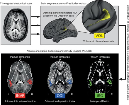
T1-weighted images were automatically segmented into gray and white matter using surface-based methods in FreeSurfer (top left). From the reconstructed cortical surface, the PT in each hemisphere was defined according to the Destrieux atlas (top right) and volume estimates (VOL) were obtained. The PT was linearly transformed into the native space of the diffusion-weighted NODDI images, and different microstructural measures (INVF, ODI, and ISO) were computed (bottom). ROIs, regions of interest.