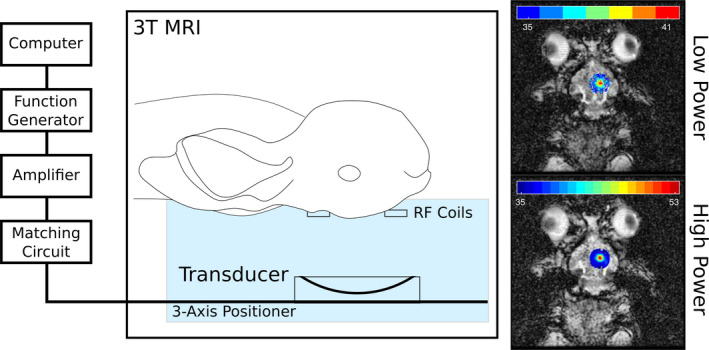
Figure 3.

The experimental setup during the in vivo rabbit experiments. The rabbit is placed supine and the transducer (f = 1.5 MHz) is positioned using a three‐axis positioner to sonicate a central target close to the surface of the brain, to avoid skull base heating. Radiofrequency (RF) coils are placed close to the target for localized thermometry with a 3 T MRI system during the treatment. Outside of the magnet room, a custom‐built computer interface is used to perform the sonication and provide thermometry feedback. Also illustrated are examples of low‐ and high‐power sonication thermometry results overlaid on anatomical MR images. [Color figure can be viewed at wileyonlinelibrary.com]
