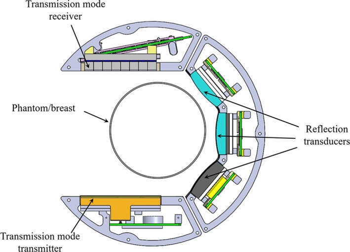
Figure 2.

Schematic of the scan head within the QT scanner. At each level, the scan head performs a full 360‐degree rotation before stepping to the next level. Within each level, the transmitter‐receiver array combination (transmission mode) and the reflection arrays time multiplex the acquisition. The three reflection transducers are of different focal length such that their depth of foci combined with rotation/translation of the scan head covers the full imaging volume. Note that there is an offset between the center of the breast and the center of the transmission arrays. The 360‐degree rotation insures that all parts of the breast, at some point, are in between the transmitter‐receiver pair and that there is indeed complete coverage of k‐space (i.e., spatial Fourier transform space), as can be most easily seen with an Ewald circle analysis. [Color figure can be viewed at http://wileyonlinelibrary.com]
