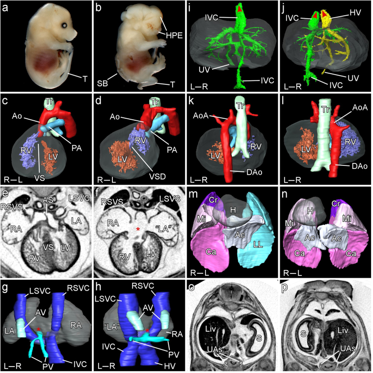
Figure 2.
Cardiovascular, pulmonary and visceral phenotypes in the Zic2 mouse mutant. Representative images illustrating range of phenotypes observed in wildtype (a,c,e,g,i,k,m,o) and homozygous mutants (iso and TALEN; b,d,f,h,j,l,n,p) at E14.5. (a,b) Holoprosencephaly (HPE), spina bifida (SB) and curly tail (T). (c,d) Reversed ventricular topology. The left ventricle (LV) is dextral to the right (RV), which gives rise to both Ao and PA (double-outlet right ventricle). (e,f) Right atrial isomerism indicated by bilateral systemic venous sinuses (SVS) and a large atrio-ventricular septal defect (asterisk). (g,h) Systemic and pulmonary veins (dorsal view). In the control embryo, the great veins (Inferior vena cava (IVC), left superior vena cava (LSVC), right superior vena cava (RSVC)) converge to form the systemic venous sinus which drains into the right atrium (RA) while the pulmonary veins (PV) drain into the left atrium (red asterisk). The azygos vein (AV) drains into the LSVC just above the level of the atria. In the mutant embryo, the IVC is left-sided while the hepatic vein (HV) and PV also drain into sinus venosus (red asterisk) which opens bilaterally into the atria. The AV is duplicated. (i,j) Hepatic venous anatomy (ventral view). In the wildtype, the IVC passes through the liver (grey shading) to drain into the right atrium (red arrowhead) and is connected to the umbilical vein (UV). In the mutant embryo, the IVC drains into the left atrium. The UV connects to the right hepatic vein (HV) which drains directly into the right atrium (red arrowheads). (k,l) Right-sided aortic arch (AoA). The descending aorta (Dao) may be seen to be ectopically located to the right of the trachea (Tr) in the mutant. (m,n) Right pulmonary isomerism. H – heart; LL – left lung; Cr – cranial lobe; Mi – middle lobe; Ca – caudal lobe; Ac – accessory lobe. (o,p) Ectopic right sided stomach (S). Liv – liver; S – stomach; UAs – umbilical arteries.

