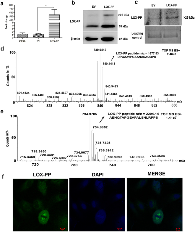
Figure 2.
Transient transfection of LOX-PP, confirmation by western blot and mass spectrometry: (a) mRNA expression of LOX-PP overexpressed in HUVECs after 48 h (b) After 48 h of transfection, cell extracts were collected and probed using LOX-PP antibody by western blot and was normalized with β- actin (EV- empty vector, LOX-PP-LOX-PP transfected cells). The corresponding full-length blots are represented in Supplementary Figs 3 and 4. (c) Conditioned medium was collected and it was immunoprecipitated using LOX-PP antibody, immunoprecipitated samples were subjected to western blot using antibody against LOX-PP and coomassie staining was considered as loading control, the corresponding full-length blots are represented in Supplementary Figs 5 and 6. (d) Mass spectrum of LOX-PP in cell lysate. (e) Mass spectrum of LOX-PP in conditioned medium. (f) Immunofluorescence for LOX-PP in HUVECs after overexpression (Magnification 63x).Cells were stained with Alexa 488 and counterstained with DAPI.

