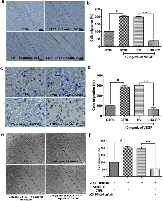
Figure 6.
Effect of LOX-PP on VEGF induced cell migration in HUVECs: (a) For scratch assay HUVEC cells were induced with 10 ng/mL of VEGF, 10 ng/mL of VEGF + EV and 10 ng/mL of VEGF + LOX-PP for 18 h and were imaged using Axio observer microscope at 5x magnification. (b) Bar graph represents the quantification of scratch assay. (c) For transwell migration assay HUVEC cells were induced with 10 ng/mL of VEGF, 10 ng/mL of VEGF + EV and 10 ng/mL of VEGF + LOX-PP for 8 h and were imaged using Axio observer microscope at 5x magnification. (d) Bar graph represents the quantification of transwell migration assay. Cells were counted using ImageJ software (expressed as % of controls). (e) HUVEC cells were induced with 10 ng/mL VEGF, 10 ng/mL VEGF + Vehicle ctrl and 10 ng/ml of VEGF + 2.5 μg/ml of rLOX-PP for 18 h and were imaged using Axio observer microscope at 5 x magnification. (f) Bar graph represents the quantification of scratch assay. Values were expressed as mean ± SD, n = 3. ***p < 0.001 versus VEGF + EV. ***p < 0.001 versus VEGF + Vehicle ctrl. #p < 0.05 versus CTRL. (CTRL – Control; EV – Empty vector; Vehicle ctrl – Vehicle control).

